At Shenoy Multi-Speciality Hospitals, our X-Ray unit is equipped with a fully digital imaging system that delivers high-resolution images with significantly lower radiation exposure than conventional film-based X-rays. Whether you are here for a routine chest X-ray or an emergency bone injury, our radiology team ensures fast, accurate results.
As one of the leading multispecialty hospitals in Marredpally, we understand that timely imaging is the foundation of an accurate diagnosis. A delayed or unclear image can delay treatment — we do not allow that to happen.
Our digital X-ray system captures images instantly, processes them without film development, and makes them immediately available to your treating doctor. In emergencies, this speed can make a critical difference to patient outcomes.
From fracture assessment and chest infections to pre-operative workups and ICU bedside imaging, our X-ray facility supports every clinical department across the hospital around the clock.
Understanding Our X-Ray Services
X-ray (radiography) is one of the oldest and most widely used diagnostic imaging techniques in medicine. It uses a small, controlled dose of ionising radiation to create images of the internal structures of the body — primarily bones, lungs, and the abdomen.
Our team provides X-ray services in Secunderabad that combine advanced digital technology with fast reporting to support accurate clinical decision-making across all specialities.
Our radiographers are trained to position patients correctly for every type of X-ray view, minimising the need for repeat exposures and ensuring diagnostic quality images on the first attempt.
Before the X-Ray
A brief preparation ensures your images come out clear and usable.
- Clinical History Review: Our radiology staff reviews the requesting doctor's clinical notes to confirm the correct imaging view and technique for your condition.
- Patient Screening: We check for pregnancy, implanted devices, and allergies before proceeding — particularly important for fluoroscopic or contrast studies.
- Patient Positioning: Our radiographers carefully position you to capture the exact anatomical view required, using lead shielding on non-target areas to minimise radiation exposure.
During the X-Ray
The procedure is quick, painless, and non-invasive.
- Image Capture: You will be asked to hold still briefly while the image is taken. For a chest X-ray, you will typically hold a deep breath for 1 to 2 seconds. The entire exposure takes a fraction of a second.
- Digital Processing: Unlike traditional film X-rays, our digital system processes the image instantly — no darkroom, no waiting. The image appears on the radiologist's workstation within seconds.
- Multiple Views When Needed: For fractures, joints, or spine studies, we may take two or more views (e.g., AP and lateral) to give the doctor a complete picture of the area.
After the X-Ray
Imaging is only as valuable as the report that follows.
- Radiologist Reporting: A qualified radiologist reviews your images and prepares a detailed written report for your treating doctor, highlighting any abnormal findings.
- Digital Storage and Sharing: All images are stored in our digital archive (PACS) and can be instantly shared across departments or printed for your records. No physical films to carry or misplace.
Types of X-Ray Studies We Offer
Our X-ray unit supports a wide range of imaging studies across all clinical specialities.
Chest X-Ray
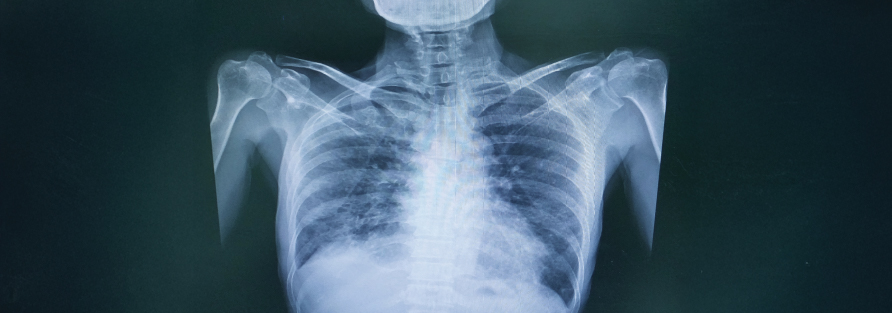
The most frequently requested X-ray in any hospital. Used to assess the lungs, heart size, ribcage, and major blood vessels. Essential for diagnosing pneumonia, tuberculosis, pleural effusion, pneumothorax, heart failure, and as a routine pre-operative check.
Bone and Joint X-Ray
Used by our orthopaedic team to assess fractures, dislocations, bone infections, arthritis, and tumours. We cover all skeletal regions — upper and lower limbs, hands, feet, pelvis, and skull.
- Fracture Assessment: Immediate X-ray for trauma patients to identify fracture type, displacement, and joint involvement — essential for deciding between conservative management and surgical intervention.
- Post-Operative Imaging: After bone fixation surgeries, X-rays confirm correct implant placement, alignment, and early signs of healing.
Spine X-Ray
Imaging of the cervical (neck), thoracic (mid-back), or lumbar (lower back) spine. Used to assess spondylosis, disc space narrowing, vertebral fractures, scoliosis, and alignment in patients with chronic back or neck pain.
Abdominal X-Ray
A plain abdominal X-ray (also called KUB — Kidney, Ureter, Bladder) is used to detect kidney stones, intestinal obstruction, bowel perforation (air under the diaphragm), and foreign body ingestion.
Dental X-Ray (OPG)
Our Orthopantomogram (OPG) provides a single panoramic image of all the teeth, the upper and lower jaw, and surrounding bone structures. Used routinely by our dental and maxillofacial surgery team for treatment planning.
Portable Bedside X-Ray
For patients in the ICU, post-operative ward, or those too unwell to be moved, our mobile X-ray unit comes to the bedside. Commonly used for daily monitoring of endotracheal tube position, central line placement, and lung status in ventilated patients.
Get Expert Diagnostic Imaging at Shenoy Hospitals
Patient Safety and Radiation Care
Concerns about radiation are common and completely valid. We take radiation safety seriously and follow the ALARA principle — As Low As Reasonably Achievable — in every imaging study we perform.
Minimising Radiation Exposure
Our digital X-ray system requires a fraction of the radiation dose compared to older film-based units while producing superior image quality. Every X-ray is performed only when clinically indicated — we do not image unnecessarily.
Protective Measures
- Lead Shielding: Lead aprons and gonad shields are used to protect body parts not being imaged, particularly for children and patients of reproductive age.
- Special Precautions for Pregnant Women: We screen all female patients of childbearing age before imaging. If pregnancy is confirmed or suspected, we discuss the clinical necessity and use abdominal shielding if the X-ray must proceed. Non-radiation alternatives like ultrasound are recommended wherever possible.
- Paediatric Protocols: Children receive lower radiation doses through adjusted imaging parameters. We also use child-friendly positioning aids to minimise the need for repeat exposures.
Cost of X-Ray Services in Secunderabad
X-ray charges at Shenoy Hospitals are straightforward and transparent. The cost varies depending on the body region and number of views required.
| X-Ray Study | Estimated Price Range (INR) |
|---|---|
| Chest X-Ray (PA View) | ₹150 – ₹300 |
| Bone / Joint X-Ray (Single Region) | ₹200 – ₹500 |
| Spine X-Ray (per region) | ₹300 – ₹600 |
| Abdominal X-Ray (KUB) | ₹200 – ₹400 |
| Dental X-Ray (OPG) | ₹400 – ₹700 |
| Portable Bedside X-Ray (ICU / Ward) | ₹400 – ₹800 |
Why do costs vary?
- Number of Views: Some studies require two or more projections (e.g., AP and lateral), which affects the total cost.
- Body Region: Larger or more complex anatomical areas require more precise positioning and may need additional images to fully assess the region.
- Bedside vs. Radiology Unit: Portable bedside X-rays involve mobilising equipment and additional technical support, which is reflected in the pricing.
X-ray charges are included in most inpatient surgical packages. Our billing team provides a complete cost breakdown before any procedure. We also process insurance and Aarogyasri / CGHS claims.
Why Choose Shenoy Multi-Speciality Hospitals?
Accurate imaging is the starting point of accurate treatment. Here is why patients and doctors across Secunderabad trust our X-ray facility:
- Fully Digital System: Our CR/DR digital X-ray unit eliminates film processing entirely, delivering instant, high-resolution images with lower radiation dose than conventional systems.
- Experienced Radiologists: All X-ray images are reported by qualified radiologists who provide detailed, clinically useful reports — not just image descriptions.
- 24/7 Availability: As a provider of super specialty health care in Secunderabad, our X-ray unit operates round the clock to support emergency, ICU, and surgical cases at any hour.
- In-Hospital Integration: Because our radiology unit is integrated within the hospital, images are instantly accessible to the treating specialist — no delays in referral or image transfer.
- Radiation Safety First: We follow AERB guidelines and ALARA principles for every scan, with lead shielding available and mandatory pregnancy screening protocols in place.
- Digital Records and Reports: All images and reports are stored digitally for easy retrieval at any future visit, eliminating the burden of maintaining physical films.
Conclusion
An X-ray is often the first step in understanding what is happening inside your body. At Shenoy Multi-Speciality Hospitals, we make that first step fast, safe, and accurate — with digital technology, trained radiographers, and expert radiologist reporting available around the clock.
Whether you need a quick chest X-ray before a procedure or an urgent bone assessment after an injury, our radiology team is ready to deliver results you and your doctor can rely on.
Do not delay your diagnosis. Walk in or book ahead — we are here when you need us.
Frequently Asked Questions
1. Is X-ray radiation harmful?
The radiation dose from a standard X-ray is very low and considered safe for most adults. Our digital X-ray system uses significantly less radiation than older film-based systems. The benefit of an accurate diagnosis far outweighs the minimal radiation exposure in the vast majority of cases.
2. Can pregnant women have an X-ray?
X-rays are generally avoided during pregnancy, especially in the first trimester, as radiation can potentially affect the developing foetus. If an X-ray is medically necessary, we use a lead shield to protect the abdomen. Always inform our radiographer if you are pregnant or suspect you may be — we will advise the safest option for you.
3. How long does an X-ray take?
The X-ray procedure itself takes only a few minutes. With our digital system, images are processed immediately and available for review by the radiologist without any waiting time for film development. Most patients are in and out of the X-ray room within 10 minutes.
4. Do I need to prepare for an X-ray?
Most X-rays require no special preparation. You may be asked to remove jewellery, glasses, or clothing with metal fasteners in the area being imaged. For abdominal X-rays, our team may advise fasting for a few hours beforehand. Our staff will give you clear instructions when you arrive.
5. How soon will I get my X-ray report?
With our digital X-ray system, images are available immediately after the scan. Reports from our radiologist are typically ready within a few hours for routine cases. For emergency patients, urgent reporting is prioritised and can be delivered within minutes when required.